Pregnancy
We individualise each
case according to
risk factors or
maternal-fetal pathologies
PREGNANCY
We individualise each case according to risk factors or
maternal-fetal pathologies.
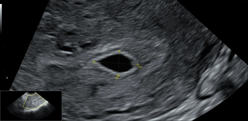
5-6 WEEKS
During this initial stage, pregnancy and fetal viability are confirmed via ultrasound. The location of the pregnancy is verified to rule out an ectopic pregnancy, and it is also determined whether the pregnancy is singleton or multiple. In addition, an initial assessment of embryonic development and the early progression of the pregnancy is performed.
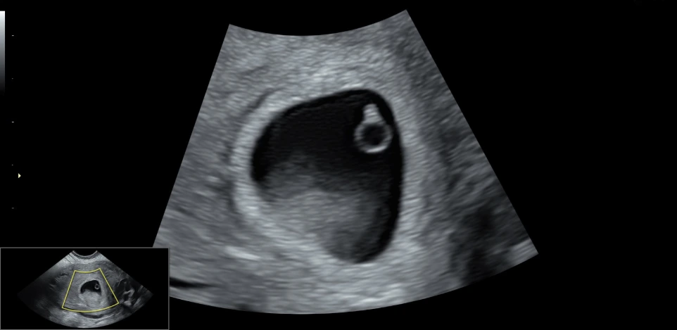
6-7 WEEKS
One of the most exciting moments of pregnancy arrives, seeing and hearing the fetal heartbeat for the first time. This ultrasound confirms embryonic cardiac activity and checks that the pregnancy is progressing correctly during the first weeks.
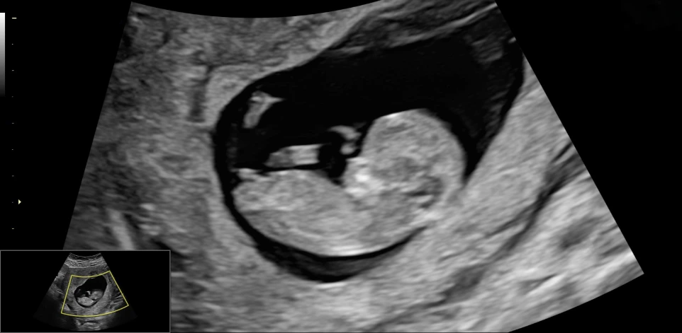
7-11 WEEKS
During this period, the growth of the embryo and its first movements begin to be clearly appreciated. Monitoring of gestational evolution is carried out, and it is the ideal time to request the non-invasive prenatal blood test for the study of chromosomal and genetic alterations.
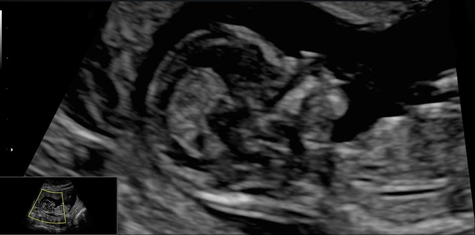
12 WEEKS
The diagnostic ultrasound of the first trimester is performed, essential for assessing fetal development. It allows for precise calculation of gestational weeks and evaluation of various ultrasound markers related to the risk of Down's syndrome and other alterations. The Doppler of uterine arteries is also studied for early detection of the risk of preeclampsia.
15-19 WEEKS
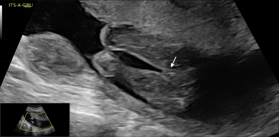
At this stage, it is already possible to identify the fetal sex, as the genitals are fully formed. Additionally, an early morphological ultrasound can be performed to assess the correct anatomical development of the baby and detect possible structural anomalies.
20-22 WEEKS
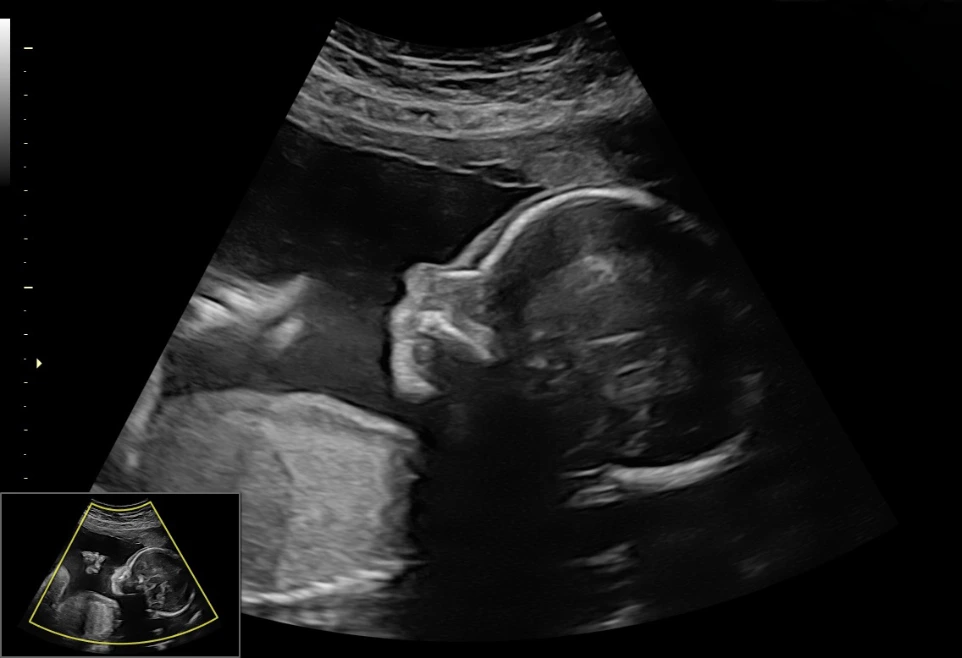
The diagnostic ultrasound of the second trimester is carried out, considered the most important ultrasound of the pregnancy. During this examination, a complete anatomical study is conducted, reviewing the fetal development organ by organ to assess growth in detail and detect possible structural alterations.
23 WEEKS THROUGH THE END OF PREGNANCY
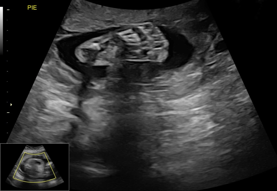
From this stage onwards, periodic ultrasound checks are carried out to assess fetal growth and well-being. The amniotic fluid, placenta, and blood flow are studied using Doppler, in addition to monitoring the baby's development during the final stretch of pregnancy.